Treatment of Uterine Fibroids with Interventional Radiology
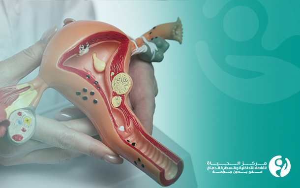
Uterine fibroids are usually benign tumors that develop in the muscular wall of the uterus. Although fibroids are often noncancerous and not life-threatening, some cases can cause troublesome symptoms and affect a patient's quality of life, such as heavy bleeding, pelvic pain, uterine enlargement, or infertility. Interventional radiology is one of the innovative treatment options for uterine fibroids and has shown promising efficacy in this field, creating real opportunities to preserve the uterus. Let's learn more about the success of treating uterine fibroids with interventional radiology.
Potential complications of uterine fibroids:
1. Pain and discomfort: Uterine fibroids can cause pain in different parts, especially if the tumors are large or close to nerves or sensitive tissues.
2. Heavy bleeding: In some cases, uterine fibroids can cause heavy bleeding, particularly during the menstrual cycle, leading to anemia and the need for treatment.
3. Pressure on nearby organs: If large fibroids grow and put pressure on neighboring organs like the bladder or intestines, they can cause problems with the function of these organs.
4. Impact on fertility: In some cases, uterine fibroids may affect fertility by interfering with the function of the uterus or fallopian tubes.
5. Pregnancy complications: Uterine fibroids can be an obstacle to pregnancy or increase the risk of miscarriage or other problems during pregnancy and childbirth.
6. Impact on daily life: Uterine fibroids may affect a woman's daily life due to pain and other annoying symptoms.
7. Potential for malignant transformation: Although uterine fibroids rarely become cancerous, they can transform into malignant tumors.
How is interventional radiology used in cases of uterine fibroids?
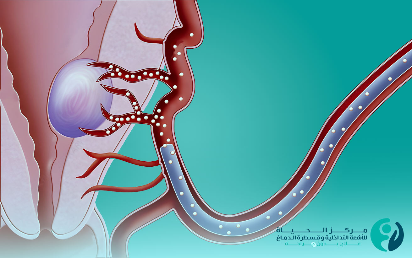
Interventional radiology treats uterine fibroids by precisely directing tiny embolic particles into the artery of the fibroid tumor to block the blood flow that nourishes the tumor, causing it to shrink in size. This treatment is performed at Al Hayat Center for Interventional Radiology and Neurointervention in Iraq using advanced techniques to ensure the accurately guidance of the catheter to the target tumor without causing significant damage to the surrounding healthy tissues.
Benefits of treating uterine fibroids with interventional radiology:
1. Non-surgical: Interventional radiology treatment is a non-surgical approach that does not require open surgery or surgical incisions. Therefore, it reduces the potential surgical complications and speeds up recovery.
2. High precision: The techniques used in interventional radiology are very accurate in injecting tiny embolic particles into the tumor blood vessel without affecting the surrounding vessels. Medical imaging like X-rays and CT scans guide the catheter accurately to the target artery.
3. Minimal impact on healthy tissue: Interventional radiology targets the fibroid tumor more than surrounding healthy tissues. Thus, it reduces the harm to healthy tissues and preserves normal uterine function.
4. No need for general anesthesia: The treatment is performed under local anesthesia, without requiring general anesthesia. This reduces the risks associated with general anesthesia and makes the treatment suitable for patients unable to tolerate general anesthesia.
5. High effectiveness: Research studies have shown the efficacy of interventional radiology in treating uterine fibroids, with the blockage of specific arteries leading to the destruction and shrinkage of tumors.
6. Avoid severe side effects: Compared to the surgery, interventional radiology is a choice that causes fewer side effects. Although some patients may experience temporary redness or pain in the treatment area, it is usually mild and temporary.
7. Preserve the uterus without removal: Interventional radiology allows for the treatment of uterine fibroids without resorting to hysterectomy and permanent loss of the hope of motherhood. Interventional radiology treatment restores the ability to conceive and complete a pregnancy peacefully.
What are the steps before starting interventional radiology treatment?
There are a few steps before starting interventional radiology treatment, which includes medical evaluation, accurate diagnosis of the condition, and necessary treatment planning. Here are some basic steps:
1. Consultation with a specialist physician: It is recommended to speak to a physician specialized in uterine fibroids treatment with interventional radiology provided by Al Hayat Center for Interventional Radiology and Neurointervention, which has the best physicians in Iraq. The physician will evaluate your condition and guide you on the required procedures.
2. Accurate diagnosis: Before treatment, it is necessary to accurately determine the type, size, and location of the fibroid tumor. This step may include examinations such as X-rays, advanced CT scans, and MRI to obtain detailed tumor images and determine the extent of its spread.
3. Session preparation: Before the session, the patient may make some preparatory measures that include avoiding certain medications, fasting for a certain period, or wearing special clothing during the session.
4. Treatment planning: The treatment sessions are determined based on the size and location of the tumor. All the treatment details are disscused with at Al Hayat Center for Interventional Radiology and Neurointervention.
The basic steps in the treatment of uterine fibroids with interventional radiology:
1. Evaluation and imaging: The patient's medical condition is evaluated and the uterine fibroids are imaged using ultrasound or magnetic resonance imaging (MRI) to determine the size and location of the tumors. This assessment aims to identify the target blood vessels for treatment.
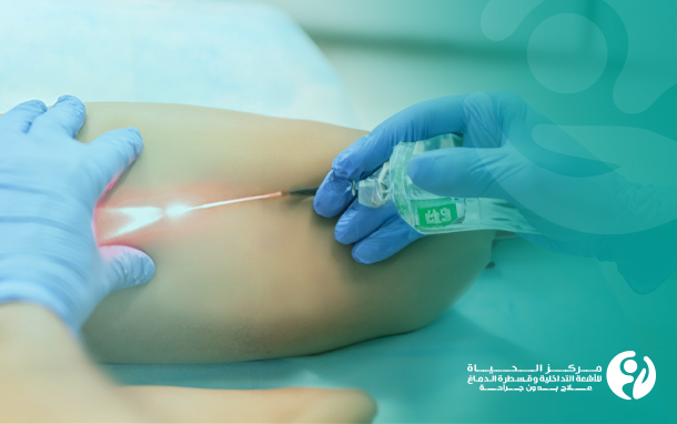
2. Local anesthesia: The patient is given a local anesthetic to minimize pain and discomfort during the procedure.
3. Catheter insertion: A thin tube known as a catheter is inserted through the femoral artery to reach the main artery that supplies blood to the uterine fibroid.
4. Interventional radiology treatment: The catheter is guided to the pelvic arteries using imaging techniques such as computed tomography (CT) or magnetic resonance imaging (MRI). Special particles are injected to block the blood supply to the tumor, leading to tissue damage and fibroid size reduction.
5. Treatment monitoring: The therapeutic effects of interventional radiology are monitored using medical imaging to verify the reduction in fibroid size and the patient's improved condition.
6. Consultation and follow-up: After the interventional radiology treatment, the patient requires follow-up visits with the specialized medical team at Al Hayat Center for Interventional Radiology and Neurointervention to verify the effectiveness of the treatment and address any potential issues.
If you are suffering from uterine fibroids and their debilitating symptoms, seek Al Hayat Center for Interventional Radiology and Neurointervention team to inquire about all the necessary steps before starting the interventional radiology treatment. They will guide you properly to ensure the safety and efficacy of the treatment.
In conclusion, interventional radiology at Al Hayat Center for Interventional Radiology and Neurointervention is a promising option for uterine fibroids treatment, as it offers numerous benefits, such as preserving the uterus, avoiding surgery, and providing high precision.